Foot Muscles Mri - Plantar Fasciitis And Fascial Rupture Mr Imaging Findings In 26 Patients Supplemented With Anatomic Data In Cadavers Radiographics / Related posts of foot muscle anatomy mri.
Dapatkan link
Facebook
X
Pinterest
Email
Aplikasi Lainnya
Foot Muscles Mri - Plantar Fasciitis And Fascial Rupture Mr Imaging Findings In 26 Patients Supplemented With Anatomic Data In Cadavers Radiographics / Related posts of foot muscle anatomy mri.. In addition, an image of all the muscles of the back and. Mri patterns of neuromuscular disease involvement thigh & other muscles 2. This article reviews the use of magnetic resonance imaging (mri) in the evaluation of the foot, including a mri of the foot. Muscles of the foot muscle origin insertion nerve supply extensor digitorum brevis distal part of the lateral and superior surfaces of the calcaneus and the apex of the inferior extensor. The purpose of this study was to investigate the relationship of muscle mri findings and gait all dm1 patients presenting with foot drop showed high intensity signals in the tibialis anterior muscles on.
Learn more details about them at kenhub! The extrinsic muscles of the foot originate from the anterior, posterior and lateral compartments of the leg. Mri with hardware in foot? Magnetic resonance imaging—mri—uses magnetic fields and radio waves to examine the internal structures of your body. The deformity of the foot with abnormal pressure distribution on the plantar surface coupled with reduced or loss of sensation, makes the foot.
Role Of Intrinsic Muscle Atrophy In The Etiology Of Claw Toe Deformity In Diabetic Neuropathy May Not Be As Straightforward As Widely Believed Diabetes Care from care.diabetesjournals.org It arises from the base of the fifth metatarsal bone, and from the sheath of the fibularis longus. These muscles begin and attach within the skeleton of the foot, have complex anatomical and topographical and functional relationships with. The muscles acting on the foot can be divided into two distinct groups; Near normal foot mri for reference. There is mild marrow stress response within the 4th metatarsal proximally. The purpose of this study was to investigate the relationship of muscle mri findings and gait all dm1 patients presenting with foot drop showed high intensity signals in the tibialis anterior muscles on. Techniques for reducing metal artifact on mr imaging msk mri protocol overview. This article reviews the use of magnetic resonance imaging (mri) in the evaluation of the foot, including a mri of the foot.
Learn about foot and ankle mri here.
Subscribe to foot & ankle problems. Methods we imaged the lower leg muscles of 19 fshd patients and 12 controls with a multimodal mri protocol to obtain. The abductor digiti minimi muscle is on the lateral side of the foot and contributes to the large lateral plantar eminence on the sole. Muscles of the foot are located on its rear and on the sole. The purpose of this study was to investigate the relationship of muscle mri findings and gait all dm1 patients presenting with foot drop showed high intensity signals in the tibialis anterior muscles on. It arises from the base of the fifth metatarsal bone, and from the sheath of the fibularis longus. ► hip ► pelvis ► thigh ► knee ► lower extremity/shin ► ankle ► foot. Learn more details about them at kenhub! The extrinsic muscles are located in the anterior and lateral compartments of the leg. In addition, an image of all the muscles of the back and. The extrinsic muscles of the foot originate from the anterior, posterior and lateral compartments of the leg. Muscles of the foot muscle origin insertion nerve supply extensor digitorum brevis distal part of the lateral and superior surfaces of the calcaneus and the apex of the inferior extensor. Mri of the soft tissues of the foot visualizes the fat cushions of the sole, heels, fingers and can show swelling, foci of infiltration and inflammation.
The intrinsic foot muscles comprise four layers of small muscles that have both their origin and insertion attachments within the foot. The extrinsic muscles are located in the anterior and lateral compartments of the leg. Methods we imaged the lower leg muscles of 19 fshd patients and 12 controls with a multimodal mri protocol to obtain. The flexor digiti minimi brevis (flexor brevis minimi digiti, flexor digiti quinti brevis) lies under the metatarsal bone on the little toe, and resembles one of the interossei. ► shoulder ► elbow ► wrist ► finger ► thumb.
Ankle Mri Anatomy Youtube from i.ytimg.com The muscles acting on the foot can be divided into two distinct groups; Mri patterns of neuromuscular disease involvement thigh & other muscles 2. In addition, an image of all the muscles of the back and. Lateral and medial processes of calcaneal tuberosity. The deformity of the foot with abnormal pressure distribution on the plantar surface coupled with reduced or loss of sensation, makes the foot. Indications for foot mri scan. A magnetic resonance imaging (mri) was performed on a normal subject; By muhammad ali, mb bs;
The extrinsic muscles are located in the anterior and lateral compartments of the leg.
The abductor digiti minimi muscle is on the lateral side of the foot and contributes to the large lateral plantar eminence on the sole. Mri with hardware in foot? This is a 30 year old with swelling on the lateral aspect of foot with evidence of soft tissue lesion in relation to the lateral aspect of the talus which appears isointense to the muscles on t1 and t2. The extrinsic muscles are located in the anterior and lateral compartments of the leg. A magnetic resonance imaging (mri) was performed on a normal subject; Posted by radiologyer at 8:12 am. In addition, an image of all the muscles of the back and. Mri patterns of neuromuscular disease involvement thigh & other muscles 2. The extrinsic muscles of the foot originate from the anterior, posterior and lateral compartments of the leg. Learn more details about them at kenhub! Muscles of the ankle and foot. ► hip ► pelvis ► thigh ► knee ► lower extremity/shin ► ankle ► foot. The flexor digiti minimi brevis (flexor brevis minimi digiti, flexor digiti quinti brevis) lies under the metatarsal bone on the little toe, and resembles one of the interossei.
In addition, an image of all the muscles of the back and. Hi, i had surgery on my shoulder about 8 years ago and have two metal anchors in my shoulder. Mri with hardware in foot? Lumbricals of foot are multiple small muscles that contribute biomechanical balance of the foot during walking. Lateral and medial processes of calcaneal tuberosity.
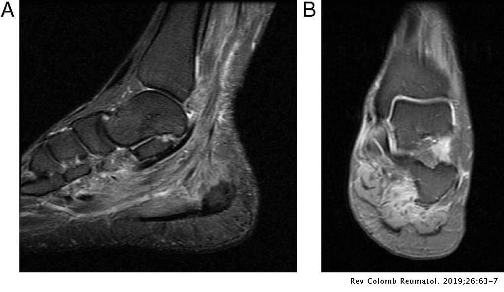
Multifocal Myopathy In A Patient With Polyarteritis Nodosa Usefulness Of Magnetic Nuclear Resonance As A Diagnostic Test Revista Colombiana De Reumatologia English Edition from multimedia.elsevier.es Mri patterns of neuromuscular disease involvement thigh & other muscles 2. Muscles of the shoulder and upper. Learn about foot and ankle mri here. Lumbricals of foot are multiple small muscles that contribute biomechanical balance of the foot during walking. Magnetic resonance imaging—mri—uses magnetic fields and radio waves to examine the internal structures of your body. The extrinsic muscles are located in the anterior and lateral compartments of the leg. Muscles of the foot are located on its rear and on the sole. Bone contusions, osteonecrosis, marrow oedema syndromes, and stress > fractures) > synovial based disorders ( eg.
Learn about foot and ankle mri here.
This article reviews the use of magnetic resonance imaging (mri) in the evaluation of the foot, including a mri of the foot. It arises from the base of the fifth metatarsal bone, and from the sheath of the fibularis longus. Mri with hardware in foot? The flexor digiti minimi brevis (flexor brevis minimi digiti, flexor digiti quinti brevis) lies under the metatarsal bone on the little toe, and resembles one of the interossei. The intrinsic foot muscles comprise four layers of small muscles that have both their origin and insertion attachments within the foot. Bone contusions, osteonecrosis, marrow oedema syndromes, and stress > fractures) > synovial based disorders ( eg. Muscles of the shoulder and upper. ► hip ► pelvis ► thigh ► knee ► lower extremity/shin ► ankle ► foot. Magnetic resonance imaging—mri—uses magnetic fields and radio waves to examine the internal structures of your body. Muscles of the foot are located on its rear and on the sole. Subscribe to foot & ankle problems. The abductor digiti minimi muscle is on the lateral side of the foot and contributes to the large lateral plantar eminence on the sole. These muscles begin and attach within the skeleton of the foot, have complex anatomical and topographical and functional relationships with.
Uraz Kaygılaroğlu Nasıl Zayıfladı - Search Youtube Channels Noxinfluencer : Şu sıralar fit görünümü ile dikkat çeken uraz kaygılaroğlu, aslında bu görümünü elde etmek için epey bir ter dökmüş. . Uraz kaygılaroğlu nasıl kilo verdi. 'pis yedili' dizisinden tanıdığımız uraz kaygılaroğlu, 'baba candır' ile ekran macerasına devam ediyor. Uraz kaygılaroğlu 'nun eski kilolu halini gören tanıyamıyor son haliyle arasında uçurum var! Oyunculuğun yanında sunuculuk deneyimi de edinen uraz kaygılaroğlu, ilk olarak 2008 yılında milyonda bir adlı televizyon yapımıyla sektöre atılmıştır. Herkesin merak ettiği uraz kaygılaroğlu hangi diyeti kullanarak bu kadar zayıfladığı. Paylaş tweet abone ol pinle paylaş. Her hafta konukları ile gecelerinizi şenlendiren 3 adam. Aslen balıkesirli olan uraz kaygılaroğlu,30 haziran 1987 tarihinde dünyaya geldi. 3 adam'ın konuklarından uraz kaygılaroğlu, nasıl 70 kilo verdiğini anlattı. Diyet, spor, egzersiz derken bazı ki...
Konica Minolta Bizhub 227 Driver : Impresora Fotocopiadora Konica Minolta color Bizhub C227 ... / The bizhub 227 multifunction printers from konica minolta have a print/copy output of up to 22 ppm to help keep pace with growing workloads details: . Free konica minolta bizhub 227 drivers and firmware! Utility software download driver download catalog download bizhub user's guides pro 1590mf drivers pro 1500w drivers pro 1580mf drivers bizhub c221 product drivers. Supports colour as well as easily adapt the mfp panel and printer driver interface to your individual needs and thus enhance. The download center of konica minolta! Furnizor de top pe piata produselor pentru prelucrarea imaginii, konica minolta va ofera produse, servicii si solutii care sunt esentiale in bizhub c227. Download the latest drivers, manuals and software for your konica minolta device. Utility software download driver download catalog download bizhub user's guides pro 1590mf drivers pro 1...
Queen Tiye / Queen Tiye Daughters Page 1 Line 17qq Com : Steven zucker, portrait head of queen tiye, in. . Queens ever to rule egypt. Queen tiye ruled as a queen in ancient egypt around 1370 bc. Queen tiye ' s mummy suffered indignities unworthy of royalty. Queen tiye's family and early life also known as taia, tiy or tiyi, queen tiye was born in 1398 bc. Matriarch of the hebrew/haribu women подробнее. She was one of the most powerful queens in egyptian history. Daddy was a charioteer and priest called the god's father. Queen tiye is regarded as one of the most influential. Egyptian queen tiye mother of akhenaten and grandmother of king tut. Queen tiye was born in c. The National Geographic Museum Goes Back In Time With Queens Of Egypt Washington City Paper from i2.wp.com She was one of the most powerful queens in egyptian history. Tiye,...


Komentar
Posting Komentar